A Word About the Circulatory System
The heart is a muscle that beats 60 to 100 times per minute. Each beat pumps oxygenated blood into the body, where it travels through a system of arteries and blood vessels. Once all organs and tissues have received nutrients and oxygen, blood returns to the heart, and then to the lungs, where it picks up more oxygen. The cycle of circulation is ongoing, with pathways that move in only one direction to ensure that oxygenated blood gets delivered and deoxygenated blood is returned to the heart. Vascular ultrasounds are designed to observe parts of this system, including arteries, veins, and capillaries.
Types of Vascular Ultrasound
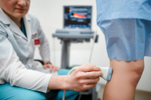
All ultrasound screenings are noninvasive. The test does not hurt because it involves the technician moving a small handpiece over the skin using very light pressure. The types of ultrasound that we may perform include:
- Arterial Doppler ultrasound. This type of imaging test may be recommended if a patient exhibits signs of poor arterial or venous circulation. This could occur in the neck, arms, or legs. Reduced blood flow could indicate a blood clot, injury to a blood vessel, or some type of arterial blockage, all of which can cause health implications. A Doppler ultrasound measures the pressure within targeted vessels and identifies how freely blood is flowing. It can help diagnose peripheral artery disease and other venous conditions.
- Carotid ultrasound. Also referred to as carotid duplex, this screening is performed on one side of the neck. This is where the carotid arteries are located. The carotid ultrasound measures blood flow through these arteries and can help identify a blockage. Here, a blockage is referred to as arterial plaque, a sticky combination of fat, calcium, and cellular debris. Arterial plaque is the cause of atherosclerosis, what we know as hardening of the arteries.
- Renal artery ultrasound. This type of ultrasound imaging evaluates the arteries that are responsible for oxygenating and nourishing the kidneys. Narrowing and blockage may occur in these arteries and contribute to renal dysfunction or hypertension.
Doctors may order a vascular ultrasound for a number of reasons, both to diagnose and to monitor various conditions. Patients who need to see a vascular specialist may likely need one of the various types of vascular ultrasound to help develop their personal treatment plan. To schedule a visit to our Waldorf, MD vascular center or for more information on our services, contact us at (301) 374-8540.