Working together with clinical oncologists, our physicians have developed procedures to treat many cancer patients and to improve their quality of life. Doctors treat most cancers with surgery, chemotherapy, radiation therapy, or some combination of these treatments, depending on the type and stage of a patient’s cancer.
The following interventional procedures for cancer are available at Metropolitan Vascular Institute.
Mediports
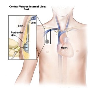
A port is a small device that is placed under the skin on your chest or arm. The port connects to a small, soft tube called a catheter which is placed inside one of the large central veins that take blood to your heart.
Patients can receive a number of medications infused through a mediport including chemotherapy. They can also be used for blood draws and transfusions, eliminating the need for multiple IV Sticks for a patient with frequent blood work and infusions. A port can stay in place for months or even years if needed.
Biopsies
We can perform a biopsy, tissue or cell sampling, of almost any organ or body part. We send our specimens out to an independent laboratory for testing. Your doctor will receive results in 2-4 days of testing. While we can provide conscious sedation for a patient’s comfort, some biopsies can be performed with just local anesthesia. Common biopsy sites are liver, bone, soft tissue, lymph node, muscle, and kidneys.
Tumor Embolization/Ablation
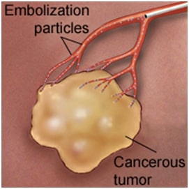
Tumor Embolization is a procedure in which small spheres are injected to try to block or reduce the blood flow to cancer cells. Embolization can be performed on many types of tumors, and/or tumors that have an abundant vascular supply. Based on the patient’s anatomy and specific treatment plan, the procedure can be performed by accessing either the femoral artery, in the groin, or the radial artery, in the wrist. All options and plans will be discussed with the physician during the patient’s initial consult.
In some cases, embolization is performed to reduce the blood supply to the tumor so that surgical resection can be performed.
Tumor Ablation
This is a minimally invasive surgical method to treat solid cancers. Special probes are used to “burn” or “freeze” cancers without the usual surgery. Computed Tomography (CT), Ultrasound (US) or Magnetic Resonance Imaging (MRI) is used to guide and position the needle probe into the tumor. This requires only a tiny hole, usually less than 3 mm via which the probe is introduced. When the probe is within the tumor it is attached to a generator which “burns” or “freezes” cancer. The effectiveness of this technique in treating cancer depends on two things:
- The size of the tumor
- Its accessibility to the probes
In general, for cancers 3 cm or less and easily accessible percutaneously the aim is to completely kill cancer. The larger the tumor the more difficult it is to achieve complete cancer death, therefore early treatment is crucial.
The most common cancers treated by this method are lung cancer, liver cancer, and kidney (renal) cancer. Other cancers can also be treated provided they are accessible and of appropriate size.
Different forms of tumor ablation include:
- Radiofrequency ablation: uses high-frequency electric current passed through a probe, causing the probe to increase in temperature killing the cancer cells.
- Cryoablation or Cryotherapy (cold): uses Argon circulated through a probe, freezing the cancer cells.
- Microwave: uses radio waves to heat the probe to kill the cancer cells.
Ablation is an effective treatment option for people who:
- Want to avoid conventional surgery
- Are too ill to undergo surgery
- Have a tumor that is too large to be removed surgically
- Have a limited number of tumors that have metastasized (spread from other parts of their body)
Tumor ablation is also effective for reducing a tumor’s size so that it can be treated more effectively by chemotherapy or radiation therapy or to improve symptoms.
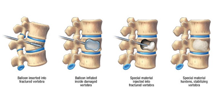
Kyphoplasty/Vertebroplasty
Kyphoplasty and vertebroplasty are used to treat painful vertebral compression fractures in the spinal column, to stabilize the bone, and to restore some or all of the lost vertebral body height due to the fracture. Such fractures are a common result of osteoporosis and can be secondary to injury, or a tumor.
During a vertebroplasty/kyphoplasty procedure, a small incision is made in the back through which the doctor places a narrow tube using imaging guidance ensuring the correct position. The tube creates a path through the back into the fractured area through the pedicle of the involved vertebrae. Your doctor may inject a cement-like material called polymethylmethacrylate (PMMA) into the fractured bone (vertebroplasty) or insert a balloon into the fractured bone to create a space and then fill it with cement (kyphoplasty). This pasty material hardens quickly, stabilizing the bone. Following vertebroplasty, about 75 percent of patients regain lost mobility and become more active.
Central Lines
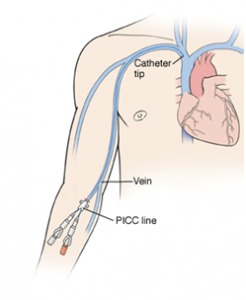
Central lines are placed for patient who may need long term blood draws and or medication injections. This allows medical personnel to administer IV medications and blood draws without multiple needle sticks in your arms. Central lines can be tunneled and non-tunneled, implanted ports, or PICC (peripherally inserted central catheter) lines. They can have 1,2, or 3 lumens, depending on why the central line is needed. The tip of the catheter is placed into the atrium of the heart with the exit site depending, as to what type of line is inserted.
Tunneled and non-tunneled central lines have exit sites at the neck, or chest wall area, while a PICC line’s exit is typically in the arm. The catheters ends have clips and caps that prevents blood from leaking out and air from entering the body.
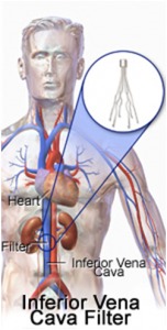
Inferior Vena Cava (IVC) Filter Placement/Removal
In an inferior vena cava filter placement procedure, interventional radiologists use image guidance to place a filter in the inferior vena cava (IVC), the large vein in the abdomen that returns blood from the lower body to the heart. The most common approach is to gain access to the jugular vein in the neck, using ultrasound guidance initially.
Blood clots that develop in the veins of the leg or pelvis, a condition called deep vein thrombosis (DVT), occasionally break up and large pieces of the clot can travel to the lungs. An IVC filter traps large clot fragments and prevents them from traveling through the vena cava vein to the heart and lungs, where they could cause severe complications or even death.
Until recently, IVC filters were available only as permanently implanted devices. Newer filters, called optionally retrievable filters, may be left in place permanently or have the option to potentially be removed from the blood vessel later. This removal may be performed when the risk of clot travelling to the lung has passed. Removal of an IVC filter eliminates any long-term risks of having the filter in place. It does not address the cause of the deep vein thrombosis or coagulation.
Your referring physician will determine if blood thinners are still necessary. However, not all retrievable IVC filters are able to be retrieved. These filters can be safely left in place as permanent filters.
Removal of an IVC Filter can be as easy as placing one. Using ultrasound guidance, the physician will access the jugular vein. In order to grab the filter, a snare or retrieval device is used to pull it out through the access sheath.
What People Say About Us!
"My experience with MVI has been a wonderful experience I have the pleasure of being the patient of Dr. Wu and Dr Gupta and their staff. They have been nothing short of a miracle. Their attentiveness and care for the patient is felt when you first meet them and I love it and the choice I made. Thank you and keep up the great work."
Click here to read more reviews.
What Can I Expect After a Tissue Biopsy?
Your doctor may request a tissue biopsy to help diagnose or rule out cancer. A biopsy may be performed on a cyst, tumor, or lump to discern what has caused it. Biopsies aren't performed only on visible bumps and lumps on or beneath the skin. Our experienced team performs this procedure on organs like the kidneys and liver, as well as bone biopsies as needed to obtain an accurate diagnosis.
What happens after your biopsy will largely depend on the type of biopsy you have done and the area from which tissue has been taken. Generally, the recovery time after a biopsy is limited to two to three days. During that time, you may take prescription or over-the-counter pain medication to address the soreness, tenderness, or pain that occurs. After a biopsy, your doctor may order other tests, such as a chest x-ray or blood count. These are done to follow-up on the biopsy and ensure that no internal bleeding has occurred.
What Should I Do and Not Do After a Tissue Biopsy?
Biopsies are carefully performed using meticulous surgical techniques. Still, your body can be stressed by the process. It's important that you rest as much as you need to for a few days following your procedure. Your doctor may also have specific activity restrictions you need to follow. For example, you may be advised to avoid using an arm or a leg if a biopsy has been performed on the limb. You may need to avoid getting the area wet for a week or two, and you'll likely be advised to avoid heavy lifting and strenuous physical activity.
How Does Kyphoplasty or Vertebroplasty Help Cancer Patients?
Some forms of cancer can spread to the bones, resulting in pain and weakening that can lead to fractures. If you have bone cancer, your doctor may advise periodic imaging or other screenings to monitor bone strength and structural integrity. Kyphoplasty and vertebroplasty are procedures that are performed when cancer has weakened one or more of the vertebrae in the spine. The procedure may be done to repair a fracture that has already occurred or to prevent a fracture in a bone that is severely weakened.
In the vertebroplasty procedure, the doctor uses a thin needle to inject bone cement into a crack. This helps restore better integrity to the bone, and it may also significantly improve comfort. According to studies, approximately 90 percent of people who undergo vertebroplasty receive remarkable pain relief. Kyphoplasty is similar to vertebroplasty but, rather than bone cement alone, inserts tiny balloons into the weakened vertebra. The balloons can increase the height of the bone before the special cement is inserted, thereby reducing pain.
The team at Metropolitan Vascular Institute is here to assist you with your questions about any of our cancer services. Please don't hesitate to contact us for further information.
