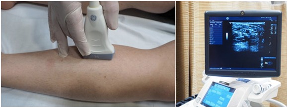
Ultrasound is a non-invasive imaging scan that uses sound waves to “see” inside your body. At the Metropolitan Vascular Institute, we have an expert team of physicians, nurses, and technologists who are highly trained in ultrasound imaging.
Venous Duplex Ultrasound Scan
The purpose of a venous duplex ultrasound scan is to evaluate venous blood flow in the patient’s arms and/or legs. Patients may experience symptoms such as pain, swelling or varicose veins in the arms or legs. These scans can aid in the diagnosis of venous abnormalities such as a suspected blood clot in a deep vein of the leg (DVT); narrowing or closure (occlusion) of a vein; or impaired blood flow (venous insufficiency).
Venous Doppler
A Venous Doppler ultrasound study may be part of a venous duplex ultrasound examination.Doppler ultrasound is a special ultrasound technique that allows the physician to see and evaluate blood flow through arteries and veins in the abdomen, arms, legs, neck, or within various body organs such as the liver or kidneys.
Renal Duplex Ultrasound Scan
The renal arteries provide blood flow to the kidneys. Renal artery disease, including narrowing (stenosis) due to atherosclerosis, can result in reduced blood-flow to the kidney. This can cause hypertension (high blood pressure). Renal artery stenosis is the most common correctable cause of hypertension. Long-standing, untreated renal artery disease is also a common cause of kidney failure.
Renal artery disease cannot be diagnosed without specific tests. Renal artery duplex scanning is accurate, non-invasive and cost-effective. Unlike angiography or CT scanning, no injection of X-ray contrast material is required, avoiding the risk of kidney damage from the contrast.
Blood-flow velocities and flow patterns in the aorta and renal arteries are evaluated with Doppler ultrasound. Imaging of the kidneys can provide information about secondary damage to the kidneys from chronic poor blood-flow.
Carotid Duplex Ultrasound Scan
A carotid duplex scan is a test that combines two types of ultrasound to look for blockages in your carotid arteries. Your carotid arteries are located along both sides of your neck. Blocked carotid arteries are a major risk factor for stroke.
Other names for a carotid duplex scan are:
- Carotid Artery Duplex
- Carotid Ultrasound
- Vascular Ultrasound
- Carotid Artery Doppler
Common Conditions That May Require Vascular Imaging
As you might imagine, vascular conditions aren't easily caught or diagnosed without a thorough imaging process. With vascular imaging, we're able to visualize and inspect blood vessels across your body for abnormalities. This allows us to more accurately assess your condition and provide more precise solutions to your vascular health concerns.
Vascular imaging can help navigate a number of conditions, including:
- Aneurysms — Vascular imaging can help identify weakened and bulging areas along blood vessel walls. It is also vital in swiftly identifying the size, location, and risk of a possible rupture.
- Varicose Veins — Though varicose veins can be identified visually on the skin, vascular imaging can help provide deeper insight into the extent of the condition.
- Deep Vein Thrombosis (DVT) — Vascular ultrasounds are a standard method for detecting the presence of blood clots characteristic of DVT.
- Peripheral Artery Disease (PAD) — This imaging can also be used successfully to evaluate the blood flow within your arteries, which helps diagnose arteries that have become narrowed or blocked due to PAD.
If the condition you're suffering from was not mentioned above, don't worry. Give our team a call here at Metropolitan Vascular Institute to learn more about how our vascular imaging in Waldorf, MD, may be able to help with your vascular condition.
Benefits of Vascular Imaging
The wide array of vascular conditions that vascular imaging can help address is just the tip of the iceberg when it comes to the benefits of this ultrasound technology. Other prominent benefits of vascular imaging and ultrasounds include:
- More accurate and timely diagnosis and intervention
- Comprehensive visualizations of your vascular network
- Clearer guidance and precision for surgical procedures
- More reliable risk assessment and prevention
- A simple, patient-friendly, and cost-effective imaging process
Our staff would be more than happy to discuss any further questions you may have about what benefits vascular imaging can provide for you.
What to Do Leading Up to Your Vascular Ultrasound Appointment
Before your vascular imaging appointment, your trusted doctor here at Metropolitan Vascular Institute will be able to provide you with clear recommendations on things you may need to do to ensure the highest quality imaging possible. Generally speaking, this often includes:
- Not eating or drinking between four and eight hours beforehand
- Ensuring that you take your medications with water
- Take an adequate dose of insulin if you are diabetic
- Avoiding any alcohol intake for the 24 hours before
Of course, these are subject to change depending on your vascular condition and the doctor's prescriptions.
When You Need Vascular Imaging, Choose Metropolitan Vascular Institute
Don't let your vascular issues go left unaddressed. If you could benefit from professional vascular imaging in Waldorf, MD, please reach out to Metropolitan Vascular Institute today. Serving the greater Waldorf area, our team can be reached online via our convenient online form or simply by giving us a call at 301-374-8540.